|

Common Eye Problems and Surgeries
Intraocular Prostheses
In many incurable glaucoma and severe trauma cases, enucleation (the removal of the eye) is necessary. In these cases, intraocular prostheses (a false eye) is an option for asthetic purposes. While the prostheses is not a functional eyeball, it will allow your pet's eye to look almost normal.
Eyelid Mass
Older dogs commonly develop eyelid tumors (cancer). As in humans, cancer can be either benign or malignant. Fortunately, eyelid tumors in dogs are usually benign and do not spread to distant tissues. However, eyelid tumors do slowly or quickly grow, and can destroy the structure of the eyelid, in addition to rubbing on the eye. It is usually best to remove them when they are still small.
Eyelid tumors are treated by surgical removal. While there are many different surgical procedures possible, some of small eyelid tumors in old dogs can be removed at Animal Eye Care without requiring general anesthesia. The patient is given a sedative, and then a local eyelid anesthetic is given to numb the eyelid. The tumor is removed and the site frozen with liquid nitrogen (cryosurgery) to kill any remaining tumor cells. Tumor cells are usually very sensitive to freezing, and normal eyelid tissue is more resistant. After surgery, the eyelid margin turns pink (depigments), but usually repigments within 4 months. When the mass is large, then a surgical removal of the surgery needs to be done under general anesthesia. Pre-operative blood test and urinalysis are required.
It is rare for the eyelid tumor to recur following surgery. 85-90% of tumors do not recur following surgery.
Cataract
A cataract is an opacity (or whitening) of the lens of the eye and may be due to old age, genetics, or diabetes. Cataracts are the major cause of blindness in dogs. Surgery is performed to remove the lens using a phacoemulsification technique (the same procedure as human cataract surgery). In some cases an artificial lens is implanted after the cataract has been removed.
Glaucoma
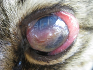
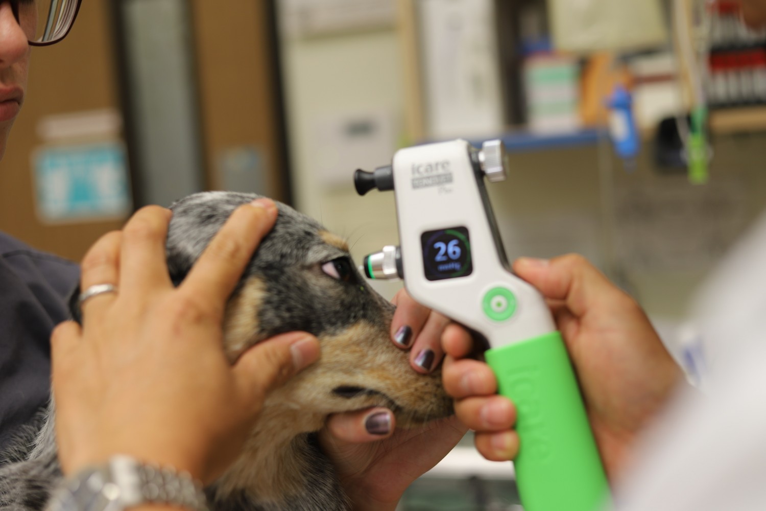
Glaucoma is an increase in pressure within the eyeball. At first the eye appears very red with a dilated pupil, then the eye becomes larger in size and the cornea looks blue or cloudy. This condition requires immediate attention and is considered a medical emergency. Medical and/or surgical attention is needed to save the sight and the eye itself. Many times the affected eye is already blind by the time the owner has recognized the condition. We use a cryotherapy (freezing) procedure and diuretics to reduce the intraocular pressure.
Cornea Ulceration and Perforations
The cornea is the clear membrane that covers the front of the eyeball. Corneal ulcerations are caused by scratches and infections of the cornea. Small, surface corneal ulcerations are not considered a medical emergency, however the deeper ulcers can cause a herniation or rupture of the Descement's membrane leading to the collapse of the eyeball. If an object penetrates to the lens, immediate surgery is needed to save the eye. Treatment options for corneal damage are different depending on the severity of damage. Chronic corneal ulcerations need special care to heal properly.
Distichiasis
Extra eyelashes appearing on the eyelid margin where they would not ordinarily grow is called distichiasis. These eyelashes are very fine and hard to notice. They often cause excessive tearing and cornea problems. Because the hair follicles are set very deep, pulling the hairs out will not solve the problem. Cryotherapy or surgery are recommended to permanently destroy the eyelashes.
Entropion
Entropion is a common eyelid abnormality where the eyelid rolls inward towards the eyeball. The most common symptoms that owners report is redness and watery eyes. Entropion is most commonly seen in shar peis, chow-chows, and rottweilers. The condition requires surgery to permanently correct the position of the eyelid.

Cherry Eye
Cherry eye is a slang term that is used to define a protrusion of the gland of the third eyelid (nictitation gland). Most of the time surgery is required to replace the gland in its proper position.
Keratitis Sicca
This condition is caused by decreased tear production. The name means dry eye and is most often mistaken as conjunctivitis. The main cause of tear gland destruction is, for the most part, unknown. However, it may be an immune mediated disease. The condition is usually permanent and requires long term treatment. Without treatment the cornea will be severely damaged and may lead to blindness and eventual enucleation (removing the eye).

Uveitis
Inflammation of the iris, cilary body, and choroid (an inflammation of the inside of the eye) is uveitis. The eye appears red and the pet tries to close its eyelids due to the pain and sensitivity to light. Uveitis can cause blindness and may require serious, long term medical treatment. This condition is seen more often in cats than dogs because of the infectious agents and diseases that they acquire (Feline Infectious Peritonitis, Toxoplasmosis, Feline Leukemia Virus, Feline Immunodeficency Virus, or fungal infections).

Progressive Retinal Atrophy
The retina is like the film in a camera. It receives an outside image and sends a picture to the brain. When the retina becomes weak and degenerated, the brain cannot receive the image and the animal becomes blind. The pupil then remains dilated and will not respond to light. This may be an inherited condition and unfortunately there is no known treatment or cure.
Feline Corneal Sequestrum
A dark, brown discoloration forms on the cornea causing pain and excessive tearing. Most of the time, surgery is required to remove the brown spots from the cornea and ocver the affected area with a conjunctival graft.
Feline Eosinophilic Keratitis
Inflammed, pink sopts appear first at the border of the cornea, then further expand toward the center of the eye and make the cat very uncomfortable. The eye at first looks like is has conjunctivitis. The condition tends to remain a long term problem and bothers the eye even with treatment.

|